
Keratoconus & Cross-Linking at Tylock George Eye Care
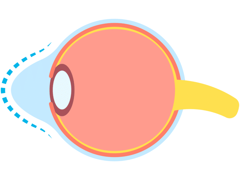
Keratoconus is a progressive eye condition in which the cornea, the eye’s clear front surface, gradually thins and bulges into a cone-like shape. This irregular shape distorts light entering the eye, leading to blurry, distorted vision and increased sensitivity to light. Often affecting people in their teens and early 20s, keratoconus can impact everyday life, making it difficult to see clearly even with glasses or contacts.
If left untreated, keratoconus can continue to progress, potentially leading to more severe vision impairment and, in advanced cases, the need for a corneal transplant. Fortunately, corneal cross-linking (CXL) is an innovative treatment designed to stop keratoconus from worsening, helping patients preserve and stabilize their vision.
What is Corneal Cross-Linking?
Corneal cross-linking is a minimally invasive procedure aimed at strengthening and stabilizing the cornea in patients with keratoconus. By reinforcing the corneal structure, cross-linking helps halt the progression of keratoconus, reducing the need for more invasive treatments in the future.
The Cross-Linking Procedure
Corneal cross-linking involves two main steps:
Application of Riboflavin Drops
Special vitamin B2 (riboflavin) eye drops are applied to the cornea, making it more responsive to the UV light used in the next step.
UV Light Exposure
After saturating the cornea with riboflavin, a controlled UV light is applied to the eye. This interaction creates new bonds between the collagen fibers in the cornea, making it stronger and more resilient.
The entire procedure typically takes around an hour, is performed on an outpatient basis, and is done under local anesthesia to ensure comfort.
Benefits of Cross-Linking
Who is a Candidate for Corneal Cross-Linking?
Ideal candidates for cross-linking:
- Has been diagnosed with keratoconus or other corneal weakening conditions
- Is experiencing progressive vision changes due to keratoconus
- Wants to preserve their vision stability and prevent further corneal deformation
Did you know, Steph Curry also has Keratoconus? You aren’t alone!
Cross-linking is most effective when performed in the early stages of keratoconus, so we recommend scheduling an evaluation as soon as possible if you suspect this condition.
Life After Cross-Linking: What to Expect
After corneal cross-linking, many patients experience increased stability in their vision. Vision may still require correction with glasses or contacts in the meantime, but the procedure helps prevent the worsening of keratoconus, creating a solid foundation for maintaining clear vision. Recovery is typically smooth, with mild discomfort and light sensitivity that resolve within a few days. If monitored by an eye doctor, it is possible to potentially review vision correction candidacy options after the Cross-Linking treatment has been completed.
Why Choose Tylock George Eye Care for Cross-Linking?
At Tylock George Eye Care, our experienced team is dedicated to helping patients with keratoconus maintain their vision and quality of life. With advanced technology and compassionate care, we guide our patients through diagnosis, treatment, and recovery to ensure the best possible outcomes. Our goal is to empower you to enjoy the highest quality vision possible, safely and effectively.
Schedule Your Consultation
If you’ve been diagnosed with keratoconus or are experiencing symptoms of distorted or blurry vision, contact Tylock George Eye Care today to schedule a consultation. We’re here to answer your questions, discuss treatment options, and help you take proactive steps to protect your vision.