The Eye Anatomy and Parts
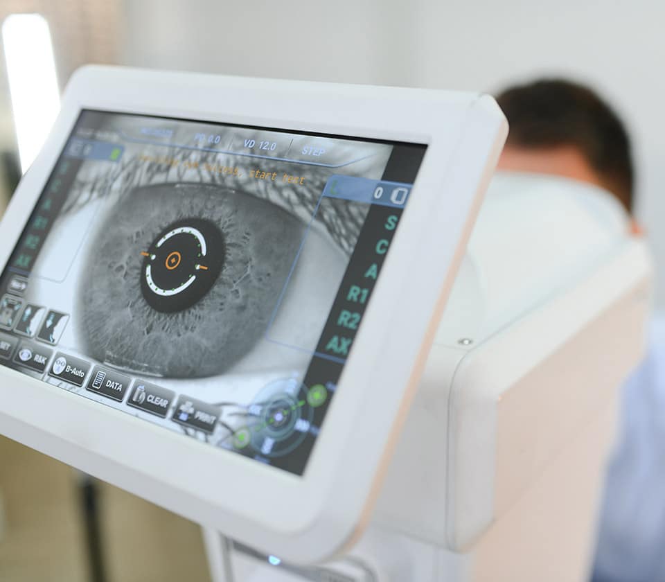
The eye is an organ of sight. With a number of components that working together for a clearer vision. Read thoroughly each eye part:

The clear dome-like structure at the front of the eye. It has about 70 percent of the refractive power or light focusing ability of the eye and acts like a fixed focus lens. Removing only a slight amount of tissue during refractive surgery has a profound effect on the way the eye sees.
The colored pigment part of the eye behind the cornea. This colored pigment covers the surface of muscles behind it that expand and contract to adjust the amount of light that enters the eye through the pupil.
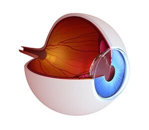
Located behind the pupil, the lens is the secondary mechanism of focus after the cornea, adjusting the amount of refraction required within the eye to focus an object on the retina. This lens becomes more and more stiff as we age, and in our 40’s causes the need for reading glasses because of its diminishing ability to focus. This condition is referred to as presbyopia.
The macula is a small area in the retina on the back wall of the eye that represents the center of vision. This area provides the clearest point of focus when viewing an object.
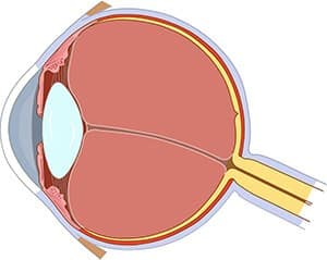
The optic nerve acts like a video cable taking visual information from the eye to the brain. Approximately 1.5 million retinal neurons converge in the back of each eye to form the optic nerve as it exits the eye. These neurons have an elaborate course as they travel through the brain terminating in the occipital cortex near the base of the skull where vision is represented.
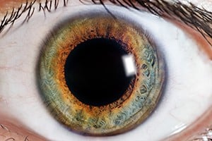
This is the black spot in the center of the eye where light enters. Pupil size changes when the iris tenses (becomes smaller) or relaxes (becomes larger) depending on the amount of light present.
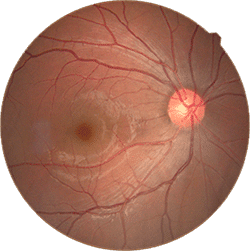
The retina is the nerve center of the eye where light is converted into an electrical signal that travels to the brain. Cells, called rods and cones, within the retina transmit these signals along the optic nerve, consequently enabling sight.

The sclera or “the white of the eye,” is the protective, opaque outer tissue of the eye. Tiny muscles connect to the sclera to control eye movement.
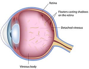
This is the gel that fills 80 percent of the eye’s volume and allows it to maintain its shape. It also serves as a clear pathway for light when it travels from the lens to the retina. The clear gel often forms clumps causing what is referred to as floaters. These floaters often have a dust or cob web appearance as they appear to float around in our vision. These floaters are not affected by refractive surgery since they are entirely inside the eye.
To learn about the function of the eye, contact Tylock Nasser Vision & Laser Center today.